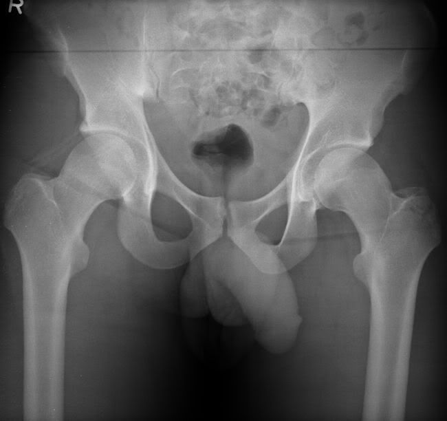
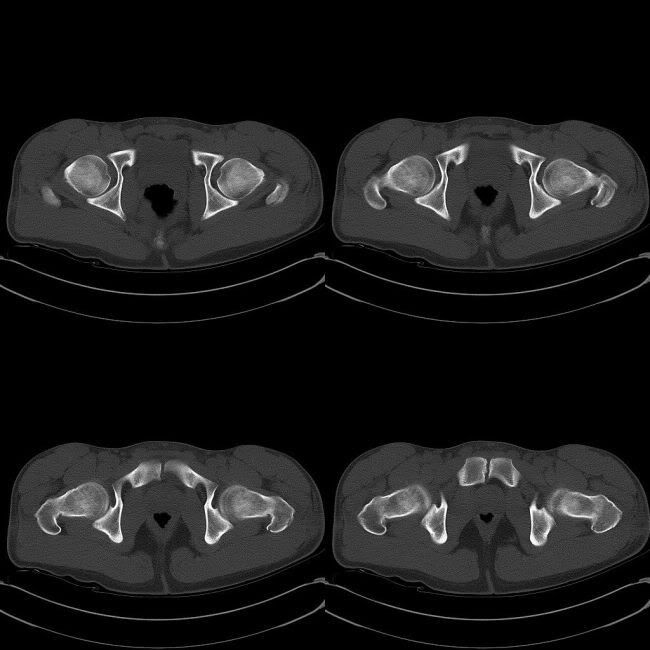
Most common site is tip and upper portion in adults. Actually more complex than what they appear on plain radiographs. MRI is useful to further characterise.
References:
Feldman F et al. MRI of Seemingly Isolated Greater Trochanteric Fractures . AJR 2004; 183:323-329
Image gallery:
Plain film:

CT correlation:
