Fibers are grouped into fascicles separated by perimysium (fibrous tissue). The whole muscle is enclosed in epimysium.
Normal ultrasound appearance:
Transverse - perimysium seen as echogenic dots or short lines scattered throughout the hypoechoic muscle fibre bulk. Intra and intermuscular septae are echogenic
Longitudinal - perimysium seen as oblique parallel echogenic stripe against hypoechoic muscle. During contraction, muscle alters the shape and is hypoechoic with increased angulation of septae
Beware of anisotropy, which results in marked hypoechogenicity, mimicing tear. To avoid this keep the transducer perpendicular to the muscle
Strains:
Grading (clinical, sonographic and MRI):
I - Spasm or cramp, stiff & sore, rapid recovery without loss of muscle strength, managed conservatively
USG - normal or focal/ generalalized) area of hyperechogenicity with or without perifascial fluid
MRI - edema/hemorrhave/both with normal muscle morphology
II - Overuse, resolve with rest, include intrasubstance tear and partial detachment of muscle from adjacent fascia or aponeurosis. Present with pain and loss of function.
USG - discontinuity of muscle fibers in echogenic perimysial striae, hypervascular, intramuscular fluid. Dynamic scanning may enhance the size and contrast of the lesion (e.g., tennis leg - medial head of gastrocnemius detaches from its common aponeurosis with soleus)
MRI - edema/hemorrhage with tear and disruption up to 50%
III - Complete myotendinous or tendoosseous tear with avulsion/retraction. Due to violent contraction against firm resistance. Early surgery may be required
USG - complete discontinuity of muscle fibers, associated hematoma. Clapper in bell sign refers to retracted echogenic muscle fragments surrounded by hypoechoic hematoma
US is superior to MR in differentiating grade 2 from 1 strains.
MRI - complete tear with retraction
Contusion:
Ill-defined hyperechogenicity in the muscle, which may cross fascial boundaries. Associated with swollen muscle.
Hematoma:
Hypoechoic fluid and may contain debris
Scar:
Hyperechoic heterogeneous, linear/stellate lesion adherent to epimysium and not change with contraction
Hernia:
Through weak aponeurosis or fascia. Hernia is better demonstrated during dynamic scan.
Myositis ossificans:
Hypoechoic mass with sheets of echogenic material and later, coarse calcifications often parallel to adjacent diaphysis
References:
1. Lee JC et al. Sonography of Lower Limb Muscle Injury. AJR 2004; 182:341-351
Image Gallery:
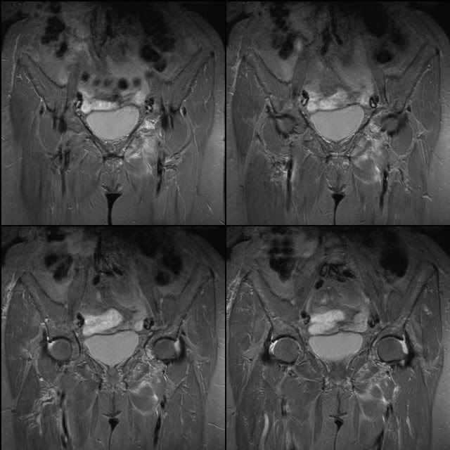
MR: Grade I muscle strain:
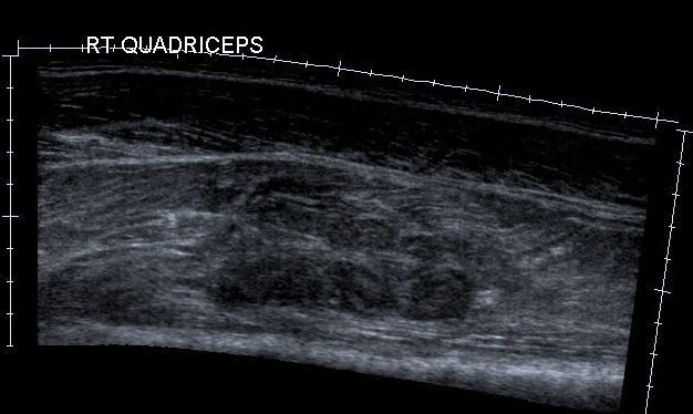
Hematoma in quadriceps: