1% of population
90% are calcaneonavicular (CN) or talocalcaneal (middle facet) (TC). CN coalition presents in childhood, where as TC present in early adulthood
Restrict subtalar eversion, inversion and anterior gliding, may cause flatfoot, pain, tarsal tunnel syndrome, tenderness, peroneal tendon spasm
Usually present in second decade of life
AD pattern is suggested
Biltaeral in about 50%
Common in rigid or spastic flatfoot
Nuclear imaging may show increased uptake
MR may show adjacent marrow edema
Usually treated conservatively with orthotics, casting, NSAID, steroid injections, physiotherapy. Surgucal options for calcaneonavicular coalition include resection with or without extensor digitorum brevis interposition, triple arthrodesis; and for talocalcaneal coalition include middle facet bony bridge resection with fat interposition, triple arthrodesis
Talocalcaneal (subtalar) coalition:
Subtalar joint consits of anterior, middle and posterior facets
AP, lateral and 45 degree internal oblique views
C sign: continuous arc between medial talar cortex and inferior cortex of sustentaculum tali on lateral radiograph. Most sensitive.
Talar beak sign: flaring of superior margin of talar head on lateral radiograph. Similar variants are hooklike osteophyte of talar head (in OA) and talar ridge (occurs more proximally)
Nonvisualized middle facet: indicated subtalar coalition on lateral view
Dysmorphic sustentaculum tali: ovoid on lateral radiograph. Normal is flat brick shaped
Other signs include: narrowing of posterior subtalar joint, rounding of lateral talar process, ball-in-socket configuration of talus
CT: coronal CT shows bony bar in middle facet of subtalar joint. In nonosseous coalition, middle facet may be narrow. Normally, sustentaculum slopes upward medially; in talocalcaneal coalition it slopes generally downward or horizontally. Sustentaculum may be broad or hypoplastic
MRI: coronal plane is best
Calcaneonavicular coalition:
Best seen on 45 degree internal oblique view. Calcaneus and navicular do not articulate.
AntEater sign: broad and elongated anterior process of calcaneus on lateral or AP radiograph. To be distinguished from normal triangular configuration of anterior process. Most sensitive sign Calcaneonavicular bar: visible bony bar or an anomalous articulation between navicular and calcaneus seen on Ap radiograph
Wide navicular: proximal articular cortex of navicular wider than articular cortex of talar head
Tapered elongated navicular: on AP view
Wide or flat anteromedial calcaneus: on AP oblique
Hypoplasia of talus: may be seen
CT:axial CT shows broadening of medial aspect of anterior and dorsal calcaneus near navicular
MRI: best on sagittal and axial MR. Sagittal images show orientation of calcaneonavicular bridging. Elongated anterior dorsal calcaneus, anteater's nose, may be seen on only one image.
References:
1. Crim JR et al. Radiographic Diagnosis of Tarsal Coalition . AJR 2004; 182:323-328
2. Joel S. Newman and Arthur H. Newberg. Congenital Tarsal Coalition: Multimodality Evaluation with Emphasis on CT and MR Imaging : RadioGraphics 2000; 20: 321
Image Gallary:
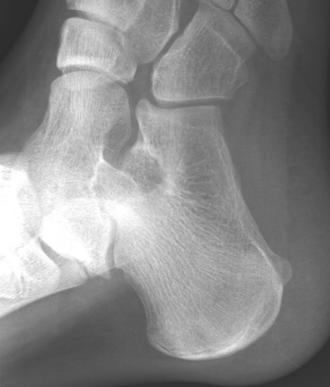
Normal lateral view: There is no C sign, no talar beak, brick shaped sustentaculum tali
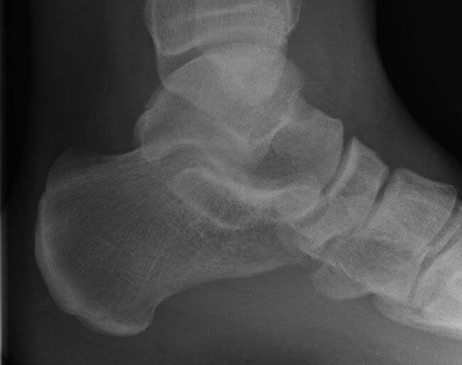
Normal foot oblique view: no 'anteater sign', no talar beak, normal middle facet, normal articular cortex of navicular