Most common inflammatory arthritis
Synovial hyperplasia and pannus formation
Affects 0.5-1% of population
Females more common than males
45-65 years
HLA DR4
Progression of disease:
Early phase: Hyperemia, early synovitis (thickening), effusion, para-articular osteoporosis, advanced synovitis (pre-erosions/subchondral cysts), erosion (starts at bare area, where bone is covered by synovium only), decreased joint space (cartilage destruction)
Late in disease: Massice erosions, bone mutilation, scarring (fibrosis), subluxation, ankylosis (bony or fibrous), intra-articular loose bodies (osseus and cartilage fragments = rice bodies)
Radiograph:
Usually normal for 6-12 months
Hyperemia is not seen
Ealry synovitis and/or effusion is seen as soft tisuue swelling
Advanced synovitis shows subchondral cysts (pre erosions) typically in bare areas
Synovitis of tendon (ECU) causes erosion of ulnar styloid process
Para-articular osteoporosis is better appreciated if compared with the previous radiograph
Joint space narrowing (due to fibrosis and destruction of cartilage) - indicated advanced stage of the disease - concentric (in OA eccentric)
Subchondral cyst, may be surrounded by sclerotic rim; tend to be large, if the joint is involved in high activity
Erosions - nearly 50% develop by 1 year
Larsen grading:
0 - normal
1 - slight changes - swelling, periarticular porosis, mild joint narrowing
2 - definite early changes - erosions, joint narrowing
3 - medium destruction - erosions in all type of joints, joint narrowing
4 - severe destruction - erosion, joint narrowing, deformity in wt bearing area
5 - mutilation - gross deformities in wt bearing areas
Steinbrocker grading:
1 - porosis
2 - 1 + muscle atrophy
3 - 2 + cartilage and bone destruction, joint deformity
4 - 3 + fibrous or bony ankylosis
Ultrasound:
Hyperemia is demonstrated on power Doppler or contrast ultrasound
Synovitis also affects tendons and extensor carpi ulnaris is early to be involved, seen on US
Effusion is readily demostrated
Erosions may be detected
Rice bodies are demonstrated
MRI:
Gd enhanced images depict hyperemia
T1 SE with early Gd (less than 5min) helps to differentiate synovium from effusion
Synovitis most commonly seen in radiocarpal joints; enhances on contrast
Gd T and T2 are good for effusions
Marrow edema (does not mean periarticular porosis) is strongly associated with subsequent erosions, subsides when erosions become inactive
Enhancement of the cortical bone
Subchondral cysts and erosions
Erosions - MR is superior to plain films; commonly seen in carpal bones, seen 4 months after the onset of symptoms; low on T1 and high signal on T2 and often enhance
Rice bodies are seen as low signal dots on T1
Differentiation from other arthropathies:
Absent proliferative changes unless secondary OA
Hall mark is bilateral symmetry with polyarticular involvement
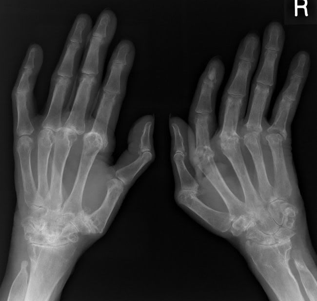
Typical - erosions of 2nd and 3rd MCPJ and PIPJ of middle finger
Wrist and hand:
Typical - erosions of 2nd and 3rd MCPJ and PIPJ of middle finger
Other joints: radiocarpal, intercarpal, MCP and PIPJ
Simultaneous tenosynovitis is common
Deformities:
Swan neck deformity - hyperextension of PIPJ and hyperflexion of DIPJ
Boutonniere deformity - flexion of PIPJ and extension of DIPJ
Hitchhiker deformity - flexion at MCPJ and extension at DIPJ
Mallet finger - drop of distal phalynges, involves medial 4 fingers
Ankle and foot:
5th MTPJ is a key target of early RA
MTPJ and IPJ are typically involved
Talonavicular, subtalar and tarsometatarsal joints are often involved than other midfoot joints
Retrocalcaneal bursitis
Glenohumaral joint:
Erosion of superolateral head
Rotator cuff tear and atrophy
Hip joint:
Not common
Asymmetry and unilateralarity are common
Erosions tends to be shallow, sclerosis milder, rarely ankylosis
Hip in rheumatoid disorders
References:
1. Sommer OJ et al. Rheumatoid Arthritis: A Practical Guide to State-of-the-Art Imaging, Image Interpretation, and Clinical Implications. RadioGraphics 2005;25:381-398
2. Sugimoto H et al. Early-Stage Rheumatoid Arthritis: Prospective Study of the Effectiveness of MR Imaging for Diagnosis. Radiology. 2000;216:569-575
3. McQueen FM. Magnetic resonance imaging in early inflammatory arthritis: what is its role? Rheumatology 2000; 39: 700-706
4. Grassi W et al. Ultrasonography in the evaluation of bone erosions. Ann Rheum Dis 2001;60:98-104 ( February )
Image gallary:
Advanced RA:
Slightly unusual RA hand:
