Tibialis anterior originates from lateral tibial condyle and proximal tibia
Musculotendinous junction is at midd third of tibia
Tendon crosses anteromedial aspect of ankle, runs towards medial border of foot
Inserts at tubercle on anteromedial medial cuneiform and inferomedial aspect of base of first metatarsal with longitudinal split, which may be visible on imaging
Superior extensor retinaculum is seen over the tendon; close to talar head superomedial band of inferior extensorretinculum; close to medial cuneiform inferomedial band of inferior extensor retinaculum and transverse retinaculum
On axial plane, it is round/oval proximally and distally with thickness of less than 5 mm
Tears:
Rarely injured or torn; hypoxic degenerative tendinosis and mucoid degeneration may cause partial or complete tear and most tears are without trauma
Present with slight foot drop, swelling and pain at dorsomedial midfoot
Common in 60- 70 years, more common
About 50 cases of complete tears are described
Located within 0.5–3 cmm from insertion
MR:
Tendon thickening of more than 5 mm and signal abnormality
Abnormal signal may be due to magic-angle effect, if the tendon is not more than 5mm
References:
1. Mengiardi B et al. Anterior Tibial Tendon Abnormalities: MR Imaging Findings. Radiology 2005;235:977-984
2. Khoury NJ et al.Rupture of the anterior tibial tendon: diagnosis by MR imaging. AJR Am J Roentgenol 1996; 167:351-354
Image Gallary:
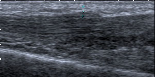
Tenosynovitis USG long axis:
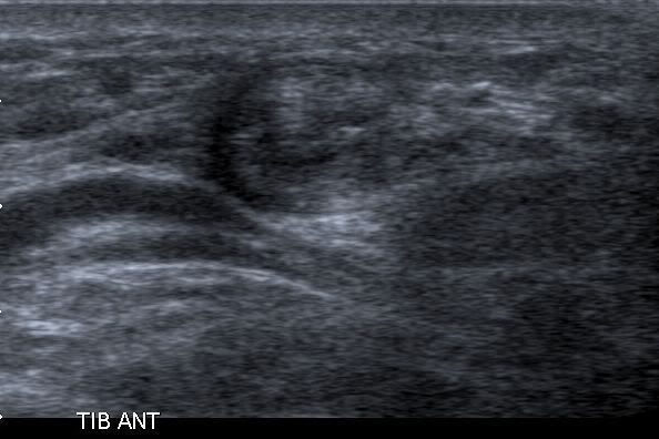
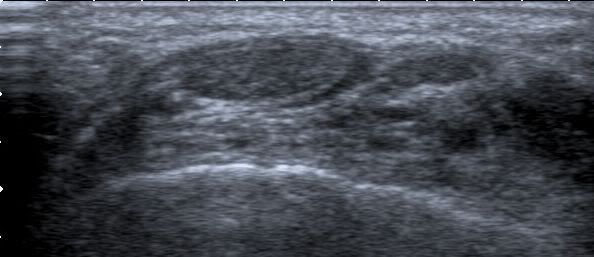
Tenosynovitis USG short axis:
Panoramic view:
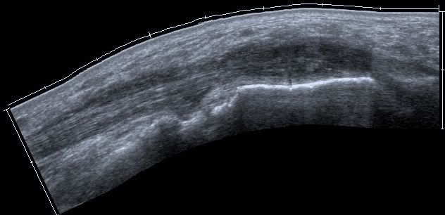
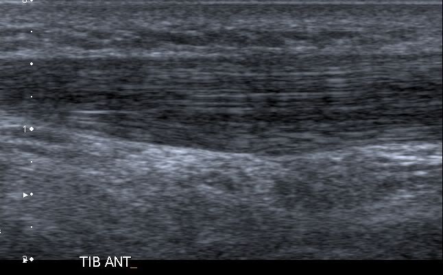
Long axis:
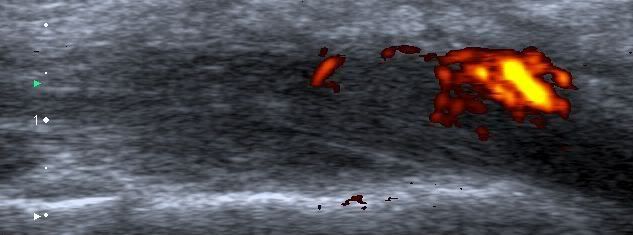
Dopppler:
Cross section: