Posterior part of the ankle joint extends into its anteroinferior corner
Retrocalcaneal bursa is situated in its posteroinferior corner
Obliterated in:
- Ruptured Achilles tendon shows soft-tissue density in Kager's fat pad, thickening of the Achilles tendon, positive Arner's sign (tendo achillis deviates anteriorly, thus nonparalleling the skin surface), decreased Toygar's angle (angle of the posterior skin surface adjacent to the tendo achillis; less than 150° is indicative of tendoachillis rupture)
- Fracture or cortical destruction of calcaneus by tumor or infection
- Os trigonum syndrome, tumours associated with FHL tendon or tendon sheath
- accessory soleus (presents as mass in the posteromedial ankle, may cause compressive neuropathy of posterior tibial nerve)
- Retrocalcaneal bursitis - posteroinferior part
- Ankle joint effusion
References:
Ly JQ et al. Anatomy of and Abnormalities Associated with Kager's Fat Pad . AJR 2004; 182:147-154
Image Gallary:
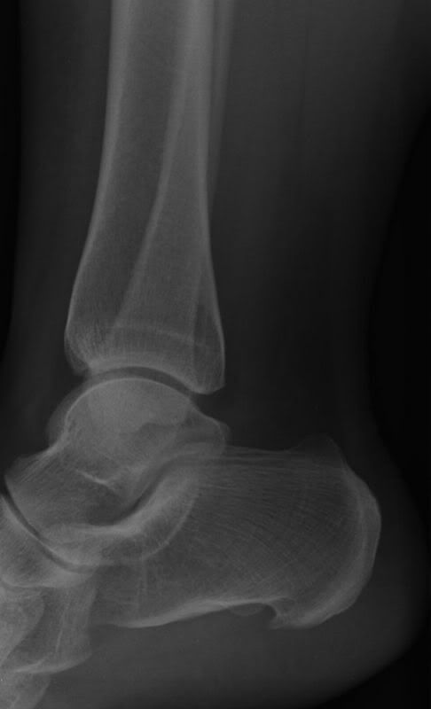
Normal Kager's fat pad: