Multilayered fibrous aponeurosis with medial, central and lateral components
Origin - medial calcaneal tuberosity
Adheres to flexor digitorum brevis
In midsole splits into five bands
Medial component covers abductor hallucis, lateral component forms investing fascia of abductor digiti minimi
Acts as strong mechanical tie for longitudinal arches by joining calcaneus, 1st metatarsal head and 5th metatarsal head
MR:
2–4-mm band
Low signal on all pulse sequences
PLANTAR FASCITIS:
Most common cause of plantar heel pain
More common on the medial side
Due to repetitive trauma or enthesopathy in association with seronegative spondyloarthropathies
More common in obese middle-aged or elderly people
Can be seen in young athletes
Pain is worse with weight bearing after a period of rest and eases with walking
Localized tenderness without swelling in anteromedial plantar surface of calcaneus
Passive dorsiflexion of toes may exacerbate pain
Radiograph:
Non-specific calcaneal spur
MR:
Fusiform proximal medial fascial thickening
Increased signal intensity
Edema of adjacent fat pad
Limited marrow edema in medial calcaneal tuberosity on STIR
Plantar fascitis and seronegative arthropathies:
Usually bilateral
Often associated with Achilles tendinitis and retrocalcaneal bursitis
More edema
More marrow changes
Management:
Conservative: weight reduction, rest, NSAID, local steroid injection, soft rubber heel pad, molded orthosis, heel cup or soft-soled shoes
No more than two local corticosteroid injections
PLANTAR FASCIA RUPTURE:
Usually sports-related injury
May be associated with local corticosteroid injection
Sudden plantar heel pain with clicking or snapping when the event occurs
Palpable tender mass
Commonly involves proximal portion at calcaneal insertion
MR:
Partial or complete interruption of fascia by high signal
Fluid around fascia
Commonly associated with underlying FDB tears
Also associated with abductor hallucis or quadratus plantae injury
Management:
Conservative - rest, shoe arch supports, orthoses, physiotherapy
PLANTAR FIBROMATOSIS:
Benign locally invasive lesion
Fibrous proliferation of plantar fascia
May be associated with other superficial fibromatoses like palmar fibromatosis
Painless nodules in plantar arch
Frequently bilateral
Usually central and medial portions of plantar fascia
MR:
Single or multiple nodular thickening of inferior margin of plantar fascia
Low-to-int on T1- and T2
XANTHOMA OF PLANTAR FASCIA:
Seen in hyperlipidemia IIa and III
Usually bilateral and symmetric
MR:
Fusiform enlargement with heterogeneous signal
Characteristic speckled or reticulated appearance on both T1- and T2-weighted images
References:
Narvaez et al. Painful heel: MR imaging findings. Radiographics: 2000: 20: 333-352
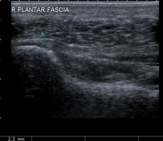
Image Gallary:
Normal plantar fascia as seen on sagittal section on ultrasound: